No products
Product successfully added to your shopping cart
There are 0 items in your cart. There is 1 item in your cart.
ELISA KIT
- Protein Control Ligand
- Pathway Inhibitors
- Enzyme Inhibitors
- Kinase Inhibitors
- Protease
- Synthase
- p18
- p38
- p53
- p70
- p90
- Peptidase
- Carboxyl and Decarboxylases
- Ceramide Turnover Enzymes
- Chromatin Modifying Enzymes
- Cyclic Nucleotide Turnover Enzymes
- Glycerophospholipid Turnover Enzymes
- Hydroxylases
- Ubiquitin-Activating Enzyme
- Adenosine Deaminase
- Clathrin
- Nuclease
- p68
- ACE
- COX
- DHFR
- Neprilysin
- NF-κB
- RAF
- RAS
- Reductase
- ROR
- Topoisomerase
- Transferase
- Protein Inhibitors
- Transporter Inhibitors
- Cell Inhibition
- Synthase
- Receptor Tyrosine Phosphatases (RTP)
- AChE
- Peptidase
- Autophagy
- Toll-Like Receptor (TLR)
- Enzyme Inhibitors
- Function Modulators
- Activators
- G Protein-Coupled Receptor Ligands
- 5HT Receptors
- Adrenoceptor
- Angiotensin Receptor
- Cannabinoid Receptors
- CCK Receptors
- DA Receptors
- EAA Receptors
- Ghrelin Receptors
- GABA Receptors
- Histamine Receptors
- Leukotriene Receptors
- Metabotropic Glutamate Receptors
- Motilin Receptors
- Muscarinic Receptor
- Neuropeptide Receptors
- Opioid Receptors
- Orexin Receptors
- Orphan Receptors
- Prostanoid Receptors
- Proteinase-Activated Receptors
- Purinergic Receptors
- Ryanodine receptor
- Sigma Receptors
- Thrombin Receptor
- Vaniloid Receptor
- VIP and PACAP Receptors
- Neurotensin Receptors
- Urotensin Receptor
- Imidazoline receptor
- SMO Receptors
- Apelin Receptor
- β-arrestin/β2-adaptin
- KDM4
- Glucocorticoid Receptor
- Laminin Receptor
- AHR
- Amylin Receptor
- Bombesin Receptor
- Bradykinin Receptor
- CFTR
- CGRP Receptor
- CRFR
- Endothelin Receptor
- Ephrin Receptor
- Farnesoid X receptor (FXR)
- Glucagon Receptor
- Nuclear Receptor Ligands
- GDNF Receptors
- TNF Receptors
- Transcription Factors
- Chemokines
- Cytokine Receptors
- Biomarkers and Buffer Solutions
- Molecular Probes
- Stem Cell Research
- Alzheimer's Disease
- Apoptosis
- Cancer Research
- Epigenetics
- Metabolites
- PET/SPECT Imaging Precursors
- Customized Screening Library
- Ultra Pure Pharmacological Standard
- Tissue Microarray (TMA)
- Proteins and Antibodies
- Primary Cells
- ELISA KIT
- Natural Products
- Lab Equipments
- Humanized Mice for PDX Platform
- Rare Chemicals
- Custom Synthesis
- Antibacterial
- Antifungal
- Antioxidant
- Antiviral
- Molecular Glues
- PROTAC Linker
- SARS-CoV
 View larger
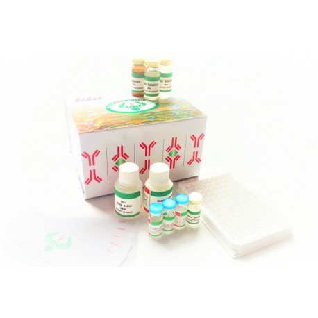
 ELISA Kit FOR Tumor necrosis factor receptor superfamily member 9
E9264h
Molarity Calculation Cart®
HOW TO ORDER
More info
Intended use
This immunoassay kit allows for the in vitro quantitative determination of human Tumor necrosis factor receptor superfamily member 9, concentrations in serum, Plasma, Urine, tissue homogenates and Cell culture supernates and Other biological fluids.
Test principle
The microtiter plate provided in this kit has been pre-coated with an antibody specific to Tumor necrosis factor receptor superfamily member 9. Standards or samples are then added to the appropriate microtiter plate wells with a biotin-conjugated polyclonal antibody preparation specific for Tumor necrosis factor receptor superfamily member 9 and Avidin conjugated to Horseradish Peroxidase (HRP) is added to each microplate well and incubated. Then a TMB substrate solution is added to each well. Only those wells that contain Tumor necrosis factor receptor superfamily member 9, biotin-conjugated antibody and enzyme-conjugated Avidin will exhibit a change in color. The enzyme-substrate reaction is terminated by the addition of a sulphuric acid solution and the color change is measured spectrophotometrically at a wavelength of 450 nm ± 2 nm. The concentration of in the samples is then determined by comparing the O.D. of the samples to the standard curve.
Materials and components
Reagent Quantity
Assay plate 1
Standard 2
Sample Diluent 1 × 20ml
Assay Diluent A 1 × 10ml
Assay Diluent B 1 × 10ml
Detection Reagent A 1 × 120μl
Detection Reagent B 1 × 120μl
Wash Buffer(25 x concentrate) 1 × 30ml
Substrate 1 × 10ml
Stop Solution 1 × 10ml
Plate sealer for 96 wells 5
Instruction 1
Other supplies required
Microplate reader
Pipettes and pipette tips
EP tube
Deionized or distilled water
Storage of the kits
The Assay Plate, Standard, Detection Reagent A and Detection Reagent B should be stored at -20℃ upon being received. After receiving the kit, Substrate should be always stored at 4℃. Other reagents are kept according to the labels on vials. But for long term storage, please keep the whole kit at -20℃. The unused strips should be kept in a sealed bag with the desiccant provided to minimize exposure to damp air. The test kit may be used throughout the expiration date of the kit (six months from the date of manufacture). Opened test kits will remain stable until the expiring date shown, provided it is stored as prescribed above.
Sample collection and storage
Serum - Use a serum separator tube (SST) and allow samples to clot for 30 minutes before centrifugation for 15 minutes at approximately 1000 × g. Remove serum and assay immediately or aliquot and store samples at -20℃ or -80℃.
Plasma - Collect plasma using EDTA or heparin as an anticoagulant. Centrifuge samples for 15 minutes at 1000 × g at 2℃ - 8℃ within 30 minutes of collection. Store samples at -20℃ or -80℃. Avoid repeated freeze-thaw cycles.
Urine - Aseptically collect the first urine of the day (mid-stream), voided directly into a sterile container. Centrifuge to remove particulate matter, assay immediately or aliquot and store at ≤ -20℃. Avoid repeated freeze-thaw cycles.
Tissue homogenates - The preparation of tissue homogenates will vary depending upon tissue type. For this assay, tissue was rinsed with 1X PBS to remove excess blood, homogenized in 20 mL of 1X PBS and stored overnight at ≤ -20℃ After two freeze-thaw cycles were performed to break the cell membranes, the homogenates were centrifuged for 5 minutes at 5000 x g. Remove the supernate and assay immediately or aliquot and store at ≤ -20℃.
Cell culture supernates and Other biological fluids - Remove particulates by centrifugation and assay immediately or aliquot and store samples at -20℃ or -80℃. Avoid repeated freeze-thaw cycles.
Note:
- Samples to be used within 5 days may be stored at 2-8℃, otherwise samples must stored at -20℃ (1 month) or -80℃ (2 months) to avoid loss of bioactivity and contamination.
- Tissue or cell extraction samples prepared by chemical lysis buffer may cause unexpected ELISA results due to the impacts of certain chemicals.
- Influenced by the factors including cell viability, cell number and also sampling time, samples from cell culture supernatant may not be detected by the kit
- Sample hemolysis will influence the result, so hemolytic specimen can not be detected.
- When performing the assay slowly bring samples to room temperature.
- Do not use heat-treated specimens.
Limitations of the procedure
- AOBIOUS is only responsible for the kit itself, but not for the samples consumed during the assay. The user should calculate the possible amount of the samples used in the whole test. Please reserve sufficient samples in advance.
- The kit should not be used beyond the expiration date on the kit label.
- Do not mix or substitute reagents with those from other lots or sources.
- If samples generate values higher than the highest standard, further dilute the samples with the Sample Diluent and repeat the assay. Any variation in standard diluent, operator, pipetting technique, washing technique,incubation time or temperature, and kit age can cause variation in binding.
Reagent preparation
Wash Buffer - If crystals have formed in the concentrate, warm to room temperature and mix gently until the crystals have completely dissolved. Dilute 30 mL of Wash Buffer Concentrate into deionized or distilled water to prepare 750 mL of Wash Buffer.
Standard - Reconstitute the Standard with 1.0 mL of Sample Diluent. This reconstitution produces a stock solution of 200 ng/mL. Allow the standard to sit for a minimum of 15 minutes with gentle agitation prior to making serial dilutions (Making serial dilution in the wells directly is not permitted). The undiluted standard serves as the high standard (200 ng/mL). The Sample Diluent serves as the zero standard (0 ng/mL).

ng/mL 200 100. 50.0 25.0 12.5 6.25 3.12 0
Detection Reagent A and B - Dilute to the working concentration using Assay Diluent A and B (1:100), respectively.
Assay procedure
Allow all reagents to reach room temperature (Please do not dissolve the reagents at 37℃ directly). All the reagents should be mixed thoroughly by gently swirling before pipetting. Avoid foaming. Keep appropriate numbers of strips for 1 experiment and remove extra strips from microtiter plate. Removed strips should be resealed and stored at 4℃ until the kits expiry date. Prepare all reagents, working standards and samples as directed in the previous sections. Please predict the concentration before assaying. If values for these are not within the range of the standard curve, users must determine the optimal sample dilutions for their particular experiments.
- Add 100 μl of Standard, Blank, or Sample per well. Cover with the Plate sealer. Incubate for 2 hours at 37℃.
- Remove the liquid of each well, don’t wash. Add 100 μl of Detection Reagent A working solution to each well. Cover with the Plate sealer. Incubate for 1 hour at 37℃. Detection Reagent A working solution may appear cloudy. Warm to room temperature and mix gently until solution appears uniform.
- Aspirate each well and wash, repeating the process three times for a total of three washes. Wash by filling each well with Wash Buffer (approximately 400 μl) using a squirt bottle, multi-channel pipette, manifold dispenser or autowasher. Complete removal of liquid at each step is essential to good performance. After the last wash, remove any remaining Wash Buffer by aspirating or decanting. Invert the plate and blot it against clean paper towels.
- Add 100 μl of Detection Reagent B working solution to each well. Cover with a new Plate sealer. Incubate for 1 hour at 37℃.
- Repeat the aspiration/wash as in step 4.
- Add 90 μl of Substrate Solution to each well. Cover with a new Plate sealer. Incubate within 15-30 minutes at 37℃. Protect from light.
- Add 50 μl of Stop Solution to each well. If color change does not appear uniform, gently tap the plate to ensure thorough mixing.
- Determine the optical density of each well at once, using a microplate reader set to 450 nm.
Note:
- Absorbance is a function of the incubation time. Therefore, prior to starting the assay it is recommended that all reagents should be freshly prepared prior to use and all required strip-wells are secured in the microtiter frame. This will ensure equal elapsed time for each pipetting step, without interruption.
- Please carefully reconstitute Standards or working Detection Reagent A and B according to the instruction, and avoid foaming and mix gently until the crystals have completely dissolved. The reconstituted Standards Detection Reagent A and B can be used only once. This assay requires pipetting of small volumes. To minimize imprecision caused by pipetting, ensure that pipettors are calibrated. It is recommended to suck more than 10μl for once pipetting.
- To ensure accurate results, proper adhesion of plate sealers during incubation steps is necessary. Do not allow wells to sit uncovered for extended periods between incubation steps. Once reagents have been added to the well strips, DO NOT let the strips DRY at any time during the assay.
- For each step in the procedure, total dispensing time for addition of reagents to the assay plate should not exceed 10 minutes.
- To avoid cross-contamination, change pipette tips between additions of each standard level, between sample additions, and between reagent additions. Also, use separate reservoirs for each reagent.
- The wash procedure is critical. Insufficient washing will result in poor precision and falsely elevated absorbance readings.
- Duplication of all standards and specimens, although not required, is recommended.
- Substrate Solution is easily contaminated. Please protect it from light.
Specificity
This assay recognizes recombinant and natural human Tumor necrosis factor receptor superfamily member 9. No significant cross-reactivity or interference was observed.
Note:
Limited by current skills and knowledge, it is impossible for us to complete the cross- reactivity detection between human and all the analogues, therefore, cross reaction may still exist.
Detection Range
3.12-200 ng/mL.
Calculation of results
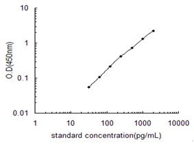
Average the duplicate readings for each standard, control, and sample and subtract the average zero standard optical density. Create a standard curve by reducing the data using computer software capable of generating a four parameter logistic (4-PL) curve-fit. As an alternative, construct a standard curve by plotting the mean absorbance for each standard on the x-axis against the concentration on the y-axis and draw a best fit curve through the points on the graph. The data may be linearized by plotting the log of the Tumor necrosis factor receptor superfamily member 9 concentrations versus the log of the O.D. and the best fit line can be determined by regression analysis. It is recommended to use some related software to do this calculation, such as curve expert 1.3. This procedure will produce an adequate but less precise fit of the data. If samples have been diluted, the concentration read from the standard curve must be multiplied by the dilution factor.
Important note:
- Limited by the current condition and scientific technology, we can't completely conduct the comprehensive identification and analysis on the raw material provided by suppliers. So there might be some qualitative and technical risks to use the kit
- The final experimental results will be closely related to validity of the products, operation skills of the end users and the experimental environments. Please make sure that sufficient samples are available.
- Kits from different batches may be a little different in detection range, sensitivity and color developing time.Please perform the experiment exactly according to the instruction attached in kit while electronic ones from our website is only for information.
- There may be some foggy substance in the wells when the plate is opened at the first time. It will not have any effect on the final assay results.
- Do not remove microtiter plate from the storage bag until needed.
- A microtiter plate reader with a bandwidth of 10nm or less and an optical density range of 0-3 OD or greater at 450nm wavelength is acceptable for use in absorbance measurement.
- Use fresh disposable pipette tips for each transfer to avoid contamination.
- Do not substitute reagents from one kit lot to another. Use only the reagents supplied by manufacturer.
- Even the same operator might get different results in two separate experiments. In order to get better reproducible results, the operation of every step in the assay should be controlled. Furthermore, a preliminary experiment before assay for each batch is recommended.
- Each kit has been strictly passed Q.C test. However, results from end users might be inconsistent with our in-house data due to some unexpected transportation conditions or different lab equipments. Intra-assay variance among kits from different batches might arise from above factors, too.
- Kits from different manufacturers for the same item might produce different results, since we haven’t compared our products with other manufacturers.
- Valid period: six months.
Precaution
The Stop Solution suggested for use with this kit is an acid solution. Wear eye, hand, face, and clothing protection when using this material.Typical Data

This graph data is shown as an example. A standard curve must be generated each time the assay is run.

