No products
Product successfully added to your shopping cart
There are 1 items in your cart. There is 1 item in your cart.
Primary Cells
- Protein Control Ligand
- Pathway Inhibitors
- Enzyme Inhibitors
- Kinase Inhibitors
- Protease
- Synthase
- p18
- p38
- p53
- p70
- p90
- Peptidase
- Carboxyl and Decarboxylases
- Ceramide Turnover Enzymes
- Chromatin Modifying Enzymes
- Cyclic Nucleotide Turnover Enzymes
- Glycerophospholipid Turnover Enzymes
- Hydroxylases
- Ubiquitin-Activating Enzyme
- Adenosine Deaminase
- Clathrin
- Nuclease
- p68
- ACE
- COX
- DHFR
- Neprilysin
- NF-κB
- RAF
- RAS
- Reductase
- ROR
- Topoisomerase
- Transferase
- Protein Inhibitors
- Transporter Inhibitors
- Cell Inhibition
- Synthase
- Receptor Tyrosine Phosphatases (RTP)
- AChE
- Peptidase
- Autophagy
- Toll-Like Receptor (TLR)
- Enzyme Inhibitors
- Function Modulators
- Activators
- G Protein-Coupled Receptor Ligands
- 5HT Receptors
- Adrenoceptor
- Angiotensin Receptor
- Cannabinoid Receptors
- CCK Receptors
- DA Receptors
- EAA Receptors
- Ghrelin Receptors
- GABA Receptors
- Histamine Receptors
- Leukotriene Receptors
- Metabotropic Glutamate Receptors
- Motilin Receptors
- Muscarinic Receptor
- Neuropeptide Receptors
- Opioid Receptors
- Orexin Receptors
- Orphan Receptors
- Prostanoid Receptors
- Proteinase-Activated Receptors
- Purinergic Receptors
- Ryanodine receptor
- Sigma Receptors
- Thrombin Receptor
- Vaniloid Receptor
- VIP and PACAP Receptors
- Neurotensin Receptors
- Urotensin Receptor
- Imidazoline receptor
- SMO Receptors
- Apelin Receptor
- β-arrestin/β2-adaptin
- KDM4
- Glucocorticoid Receptor
- Laminin Receptor
- AHR
- Amylin Receptor
- Bombesin Receptor
- Bradykinin Receptor
- CFTR
- CGRP Receptor
- CRFR
- Endothelin Receptor
- Ephrin Receptor
- Farnesoid X receptor (FXR)
- Glucagon Receptor
- Nuclear Receptor Ligands
- GDNF Receptors
- TNF Receptors
- Transcription Factors
- Chemokines
- Cytokine Receptors
- Biomarkers and Buffer Solutions
- Molecular Probes
- Stem Cell Research
- Alzheimer's Disease
- Apoptosis
- Cancer Research
- Epigenetics
- Metabolites
- PET/SPECT Imaging Precursors
- Customized Screening Library
- Ultra Pure Pharmacological Standard
- Tissue Microarray (TMA)
- Proteins and Antibodies
- Primary Cells
- ELISA KIT
- Natural Products
- Lab Equipments
- Humanized Mice for PDX Platform
- Rare Chemicals
- Custom Synthesis
- Antibacterial
- Antifungal
- Antioxidant
- Antiviral
- Molecular Glues
- PROTAC Linker
- SARS-CoV
 View larger
 Human Primary Glomerular Endothelial Cells
HUM-u003
Each vial contains >5x10⁵ cells in 1mL volume
Molarity Calculation Cart®
HOW TO ORDER
More info
Cell Details
The kidney is an organ of the vertebrate. It is part of the urinary system. It is responsible for filtering impurities in the blood, maintaining the balance between body fluids and electrolytes, and finally producing urine through the urethra. It also has endocrine function to regulate blood pressure. Observed by a microscope, it can be seen that each kidney is mainly composed of about 1 million nephrons with the same structure and function and a small amount of connective tissue, with a large number of blood vessels and nerve fibers.
Each nephron consists of a renal corpuscle and a renal tubule. The renal corpuscle has a capillary mass called the glomerulus, which is formed by the branches of the renal artery. There is a small capsular around the glomerulus. The renal capsule is divided into two layers, and there is a cyst between the two layers communicating with the lumen of the renal tubule. The renal tubules merge into a collecting tube. Several collection tubes merge into a nipple tube, and urine flows into the renal sputum.
The glomerulus is a blood filter, and the glomerular capillary wall constitutes a filter membrane. The glomerular filtration membrane has three layers from the inside to the outside: the inner layer is the endothelium, the middle layer is the glomerular basement membrane, and the outer layer is the epithelial cell layer. The blood is filtered through the filter membrane, and the filtrate is inserted into the renal capsule. Under normal circumstances, most of the protein in the blood can not be filtered and retained in the blood, only small molecules such as urea, glucose, electrolytes and some small molecules can be filtered.
Cell Characteristics
1) The cells are derived from human normal kidney tissue.
2) Cell identification: Factor VIII-related antigen (Factor VIII) was positive for immunofluorescence staining.
3) The purity of the identified cells is higher than 90% .
4) Does not contain HIV-1, HBV, HCV, mycoplasma, bacteria, yeast, and fungi.
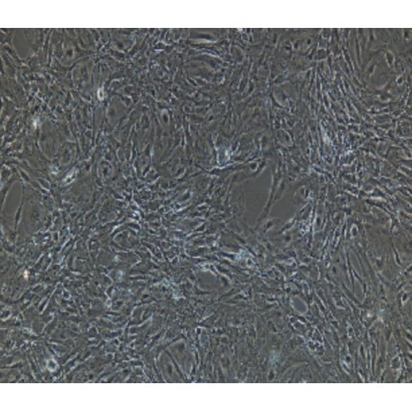
5) Cell growth mode: round, polygonal cells, adherent culture.
Transportation and Preservation
Depending on the weather conditions and the distance of transportation, the company negotiates with the customer and chooses one of the following methods.
1) 1mL of frozen cell suspension is placed in a 1.8mL cryotube and placed in a foam incubator filled with dry ice for transport; after receiving the cells, thaw the resuscitated cells as soon as possible for culture. If resuscitation is not possible immediately, Cryopreserved cells can be stored at -80°C for 1 month.
2) T-25 culture flask is filled with complete medium and then transported at room temperature. After receiving the cells, please observe the growth state of the cells under a microscope. If the bottle filling rate exceeds 85%, please carry out the subculture immediately. If there are more cells in suspension, allow the flask to stand overnight in the incubator to help the undead suspension cells to reattach.
Product Use
1) This product can only be used for scientific research
2) This product has not passed the audit for living animals and humans directly.
3) This product has not passed the audit for in vivo diagnosis.


