No products
Product successfully added to your shopping cart
There are 0 items in your cart. There is 1 item in your cart.
Primary Cells
- Protein Control Ligand
- Pathway Inhibitors
- Enzyme Inhibitors
- Kinase Inhibitors
- Protease
- Synthase
- p18
- p38
- p53
- p70
- p90
- Peptidase
- Carboxyl and Decarboxylases
- Ceramide Turnover Enzymes
- Chromatin Modifying Enzymes
- Cyclic Nucleotide Turnover Enzymes
- Glycerophospholipid Turnover Enzymes
- Hydroxylases
- Ubiquitin-Activating Enzyme
- Adenosine Deaminase
- Clathrin
- Nuclease
- p68
- ACE
- COX
- DHFR
- Neprilysin
- NF-κB
- RAF
- RAS
- Reductase
- ROR
- Topoisomerase
- Transferase
- Protein Inhibitors
- Transporter Inhibitors
- Cell Inhibition
- Synthase
- Receptor Tyrosine Phosphatases (RTP)
- AChE
- Peptidase
- Autophagy
- Toll-Like Receptor (TLR)
- Enzyme Inhibitors
- Function Modulators
- Activators
- G Protein-Coupled Receptor Ligands
- 5HT Receptors
- Adrenoceptor
- Angiotensin Receptor
- Cannabinoid Receptors
- CCK Receptors
- DA Receptors
- EAA Receptors
- Ghrelin Receptors
- GABA Receptors
- Histamine Receptors
- Leukotriene Receptors
- Metabotropic Glutamate Receptors
- Motilin Receptors
- Muscarinic Receptor
- Neuropeptide Receptors
- Opioid Receptors
- Orexin Receptors
- Orphan Receptors
- Prostanoid Receptors
- Proteinase-Activated Receptors
- Purinergic Receptors
- Ryanodine receptor
- Sigma Receptors
- Thrombin Receptor
- Vaniloid Receptor
- VIP and PACAP Receptors
- Neurotensin Receptors
- Urotensin Receptor
- Imidazoline receptor
- SMO Receptors
- Apelin Receptor
- β-arrestin/β2-adaptin
- KDM4
- Glucocorticoid Receptor
- Laminin Receptor
- AHR
- Amylin Receptor
- Bombesin Receptor
- Bradykinin Receptor
- CFTR
- CGRP Receptor
- CRFR
- Endothelin Receptor
- Ephrin Receptor
- Farnesoid X receptor (FXR)
- Glucagon Receptor
- Nuclear Receptor Ligands
- GDNF Receptors
- TNF Receptors
- Transcription Factors
- Chemokines
- Cytokine Receptors
- Biomarkers and Buffer Solutions
- Molecular Probes
- Stem Cell Research
- Alzheimer's Disease
- Apoptosis
- Cancer Research
- Epigenetics
- Metabolites
- PET/SPECT Imaging Precursors
- Customized Screening Library
- Ultra Pure Pharmacological Standard
- Tissue Microarray (TMA)
- Proteins and Antibodies
- Primary Cells
- ELISA KIT
- Natural Products
- Lab Equipments
- Humanized Mice for PDX Platform
- Rare Chemicals
- Custom Synthesis
- Antibacterial
- Antifungal
- Antioxidant
- Antiviral
- Molecular Glues
- PROTAC Linker
- SARS-CoV
 View larger
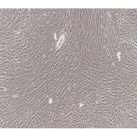
 Human Primary Bladder Smooth Muscle Cells
HUM-u008
Each vial contains >5x10⁵ cells in 1mL volume
Molarity Calculation Cart®
HOW TO ORDER
More info
Cell Details
The bladder is a urine storage organ. It is a cystic structure composed of smooth muscles. It is located in the pelvis and has a rear end opening that communicates with the urethra. There is a sphincter at the junction of the bladder and the urethra, which can control the discharge of urine.
The bladder wall is divided into three layers: the serosal layer, the muscular layer, and the mucosal layer. Smooth muscle cells are mainly in the muscle layer, and the muscle layer contains: 1. Detrusor: The detrusor is a general term for the bladder wall muscles, composed of smooth muscle. Divided into three layers, the inner and outer layers are longitudinal muscles, and the middle layer is ring muscles. The ring muscle is the thickest and strong. 2. Bladder triangle muscle: The trigone muscle is a muscle tissue other than the bladder wall layer, which starts from the ureter longitudinal muscle fibers and expands inward, downward and forward. The inwardly extending portion, and the contralateral muscles are combined with each other to form a ureteral fistula, extending downward to the posterior urethral portion, which is a Bell muscle, and a group of left and right muscle fibers intersecting at the center of the triangular region to form a triangular bottom muscle.
The contraction of the detrusor can increase the intravesical pressure and compress the urine from the urethra. There is a thick annular muscle at the junction of the bladder and the urethra to form the urethral sphincter. Syringe contraction can close the urethra to prevent urine from leaking out of the bladder.
Cell Characteristics
1) The cells are derived from human normal bladder tissue.
2) Cell identification: Smooth muscle actin (α- SMA) immunofluorescence staining was positive.
3) The purity of the identified cells is higher than 90% .
4) Does not contain HIV-1, HBV, HCV, mycoplasma, bacteria, yeast, and fungi.
5) Cell growth mode: long spindle cells, adherent culture.
Transportation and Preservation
Depending on the weather conditions and the distance of transportation, the company negotiates with the customer and chooses one of the following methods.
1) 1mL of frozen cell suspension is placed in a 1.8mL cryotube and placed in a foam incubator filled with dry ice for transport; after receiving the cells, thaw the resuscitated cells as soon as possible for culture. If resuscitation is not possible immediately, Cryopreserved cells can be stored at -80°C for 1 month.
2) T-25 culture flask is filled with complete medium and then transported at room temperature. After receiving the cells, please observe the growth state of the cells under a microscope. If the bottle filling rate exceeds 85%, please carry out the subculture immediately. If there are more cells in suspension, allow the flask to stand overnight in the incubator to help the undead suspension cells to reattach.
Product Use
1) This product can only be used for scientific research
2) This product has not passed the audit for living animals and humans directly.
3) This product has not passed the audit for in vivo diagnosis.

